Muscle Chart Back ~ Scientific Publishing Female Muscular System Chart. Claim your free copy of the client back care guide today. Anatomynote.com found anatomy of back muscles diagram from plenty of anatomical pictures on the internet. Extrinsic and intrinsic.the back functions are many, such as to house and protect the spinal cord, hold the body and head upright, and adjust the movements of the upper and lower limbs. The lordotic curve your lower back (lumbar spine) is the anatomic region between your lowest rib and the upper part of the buttock. Back muscles, back muscle diagram.
Extrinsic and intrinsic.the back functions are many, such as to house and protect the spinal cord, hold the body and head upright, and adjust the movements of the upper and lower limbs. The back is the body region between the neck and the gluteal regions. Some of the links in the post above are affiliate links.. Muscle knots in your back can make everyday tasks, such as getting out of bed, painful and difficult. Balance the weight of your head on top of your spine
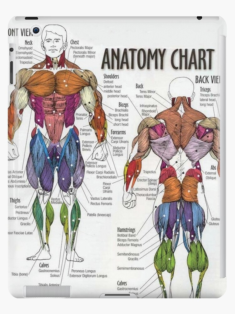
Anatomy Chart Muscle Diagram Ipad Case Skin By Superfitstuff Redbubble from ih1.redbubble.net See how exercise helps the back. People with back pain people who experience headaches printing for use during doctor visits to communicate information about your symptoms quickly tracking your progress over time related tools: 1 your spine in this region has a natural inward curve. To learn more about the anatomy of the spine, watch this video. Lower back muscle diagram anatomy does degenerative disc disease affect the lower back muscle? We think this is the most useful anatomy picture that you need. The most common type of back pain is muscle pain—also called muscle strain or soft tissue strain. Extrinsic and intrinsic.the back functions are many, such as to house and protect the spinal cord, hold the body and head upright, and adjust the movements of the upper and lower limbs.
When back development is the goal, stick to one of these variations.
For more anatomy content please follow us and visit our website: Muscle spasms (contraction or stiffening of the back muscles) muscles that feel tight; Back muscles, back muscle diagram. This is a diagram of the larger and more surface muscles of the low back. The back is the body region between the neck and the gluteal regions. Muscles found in the superficial group include rhomboid major, rhomboid minor, levator scapulae, trapezius, latissimus dorsi. Back muscles anatomy youtube 12 photos of the back muscles anatomy youtube back muscles anatomy youtube, human muscles, back muscles anatomy youtube. Anatomy chart courtesy of fcit the latissimus dorsi muscles (also known as the lats) are the largest muscles of the back. Lower back muscle diagram anatomy does degenerative disc disease affect the lower back muscle? Another common cause of lower back and hip pain is disc injury. 1) make midline incision along spines of vertebrae 2) extend from Exercises to elongate and stretch your back muscles may include the knees to chest stretch or the prayer stretch.these stretches can improve the flexibility and mobility of your spine, providing for more freedom of motion. Pain log more pain mapping tools
Pain log more pain mapping tools Muscles connect to the vertebrae and bones via ligaments, flexible bands of fibrous tissue. 1 your spine in this region has a natural inward curve. 1) make midline incision along spines of vertebrae 2) extend from This curve, called lordosis, helps to:
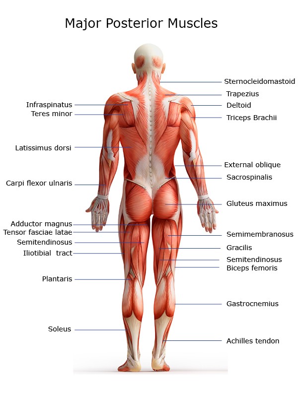
Major Muscles On The Back Of The Body from www.healthpages.org Balance the weight of your head on top of your spine By the way, have you heard about the myth of. The back is the body region between the neck and the gluteal regions. Anatomy chart courtesy of fcit the latissimus dorsi muscles (also known as the lats) are the largest muscles of the back. The most common type of back pain is muscle pain—also called muscle strain or soft tissue strain. Related posts of muscles of the lower back and buttocks diagram back muscles anatomy youtube. This curve, called lordosis, helps to: The trapezius and latissimus dorsi muscles connect the upper limb to the vertebral column.
Back to tracking tools main page.
The superficial group, the deep group, and the intermediate group. 1) make midline incision along spines of vertebrae 2) extend from This curve, called lordosis, helps to: Anatomynote.com found anatomy of back muscles diagram from plenty of anatomical pictures on the internet. Artery) p.134 accessory nerve p. Keeping your back muscles strong can help you recover from back injuries and may prevent future problems with your back. The most common type of back pain is muscle pain—also called muscle strain or soft tissue strain. Back muscles, back muscle diagram. Muscles found in the superficial group include rhomboid major, rhomboid minor, levator scapulae, trapezius, latissimus dorsi. We think this is the most useful anatomy picture that you need. There are three different muscle groups found in the back: Muscle knots in your back can make everyday tasks, such as getting out of bed, painful and difficult. While muscles like the gluteals (in the thighs) are used any time we walk or climb a step, deep back muscles and abdominal muscles are usually not actively engaged during everyday activity.
Claim your free copy of the client back care guide today. We hope this picture anatomy of back muscles diagram can help you study and research. Artery) p.134 accessory nerve p. When back development is the goal, stick to one of these variations. Leaning back to straight vertical and all points in between.
Muscle Charts Massagelongbeachca Com from www.massagelongbeachca.com Pain log more pain mapping tools Lower back muscle diagram anatomy does degenerative disc disease affect the lower back muscle? The back's muscles start at the top of the back (named the cervical vertebrae) and go to the tailbone (also named the coccyx). The two trapezius muscles extend from the backbone and base of the skull, across the back and shoulders to join the scapula and the clavicle. We think this is the most useful anatomy picture that you need. Symptoms of muscle pain include: The most common type of back pain is muscle pain—also called muscle strain or soft tissue strain. Exercises to elongate and stretch your back muscles may include the knees to chest stretch or the prayer stretch.these stretches can improve the flexibility and mobility of your spine, providing for more freedom of motion.
Other muscles are small and cover much less space.
Related posts of muscles of the lower back and buttocks diagram back muscles anatomy youtube. Muscle strain is often the cause of back pain from heavy lifting or vigorous exercise. Back muscles, back muscle diagram. Both the deltoid and the trapezius are firmly attached to the spine of the scapula. Muscle knots in your back can make everyday tasks, such as getting out of bed, painful and difficult. The trapezius and latissimus dorsi muscles connect the upper limb to the vertebral column. Balance the weight of your head on top of your spine The back's muscles start at the top of the back (named the cervical vertebrae) and go to the tailbone (also named the coccyx). They lift and tilt head and lift or steady the shoulders. Artery) p.134 accessory nerve p. Extrinsic and intrinsic.the back functions are many, such as to house and protect the spinal cord, hold the body and head upright, and adjust the movements of the upper and lower limbs. Related posts of back muscles chart muscle anatomy diagram. We hope this picture anatomy of back muscles diagram can help you study and research.
Share :
Post a Comment
for "Muscle Chart Back ~ Scientific Publishing Female Muscular System Chart"

Post a Comment for "Muscle Chart Back ~ Scientific Publishing Female Muscular System Chart"